Lion’s Mane (Hericium erinaceus) Extract 10 g – Laboratory Reference Material
- Home
- Lion’s Mane (Hericium erinaceus) Extract 10 g – Laboratory Reference Material
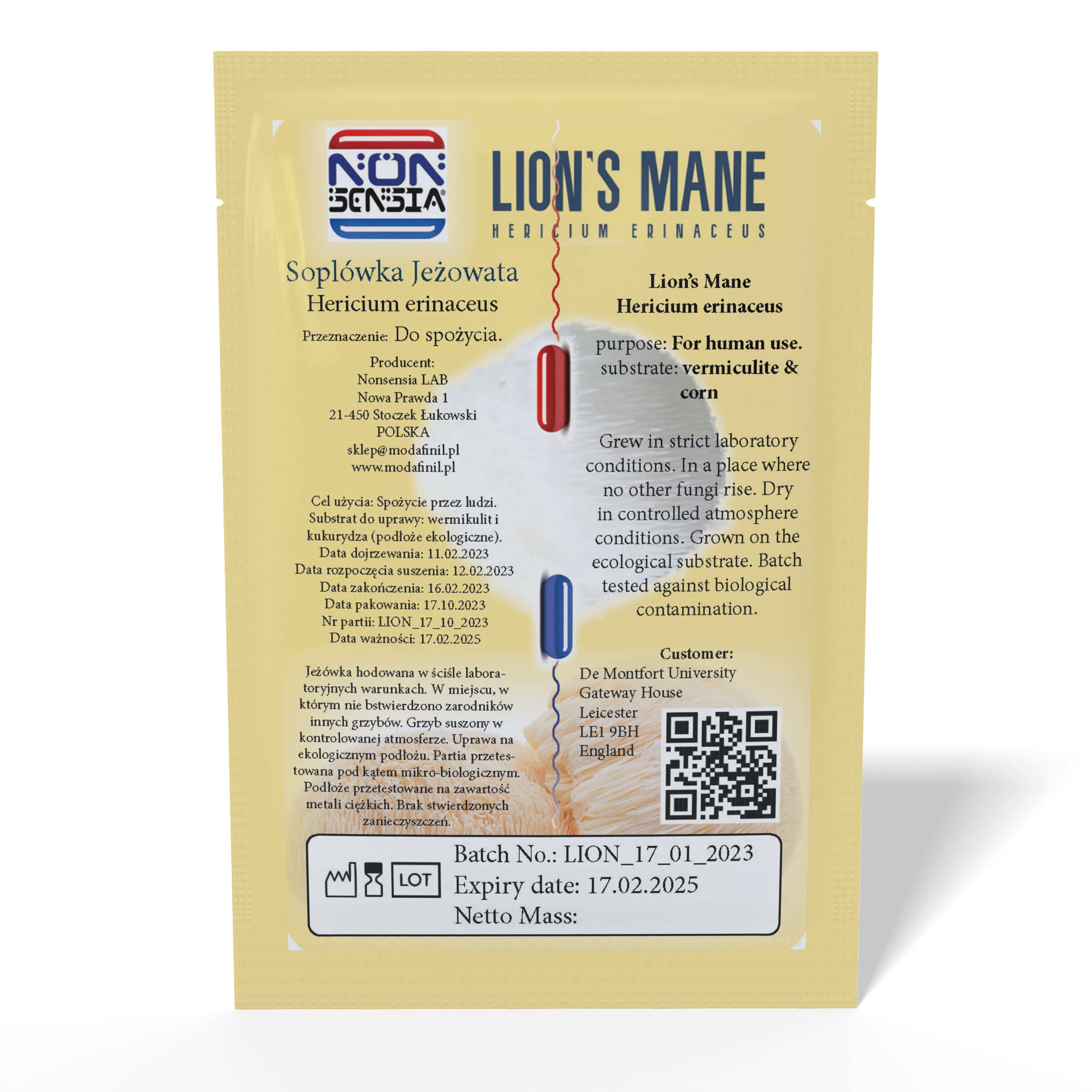
Lion’s Mane (Hericium erinaceus) Extract 10 g – Laboratory Reference Material
30,85 zł
Hericium erinaceus Extract — Laboratory Material, Marker-Defined, No Single CAS
Authentic fungal material of a stated species, supplied for extraction, fractionation and method-development work. Laboratory material only — not for human or animal consumption, and not a medicinal product, dietary supplement, food, feed or cosmetic. This is a mixture, not a compound: it has no CAS number, no molecular formula and no assignable purity, and this page explains in full what can be measured instead.
- Identity: Hericium erinaceus (Bull.) Pers. · GBIF usage key 5248508, status ACCEPTED · NCBI taxonomy ID 91752 — identifiers of an organism, not of a composition
- CAS / EC / UNII / InChIKey: none exist for this material. The European inventory returns zero records for Hericium while the same search returns EC-numbered extracts for Lentinula, Ganoderma, Grifola, Cordyceps and Trametes — the absence is real and the control fired
- Net quantity: six weighed portions, 1 g to 80 g
- What does have registry identity: thirteen marker compounds, listed with CID, CAS, formula, exact mass and InChIKey — hericenones C (137592-03-1), D, E; erinacines A (156101-08-5), C, E, S; ergosterol; ergothioneine
- The core problem: a percentage next to an extract name can mean four different quantities measured by non-equivalent procedures — named-marker content, total glucan, beta-glucan by difference, or a colorimetric index — and published data show the two common glucan methods disagreeing on both magnitude and species ranking
- Mass-spectrometric blind spot: erinacine A and erinacine E share the formula C25H36O6 and the monoisotopic mass 432.25118886 Da to the final decimal. No resolution separates them; only chromatography does
- Detector blind spot: hericenones C, D and E share one chromophore and differ only in the acyl chain — palmitate, stearate, linoleate. Ultraviolet detection cannot tell them apart, and their melting points sit 3 °C apart
- Stereochemistry, quoted verbatim: erinacine A reads 7 defined / 0 undefined atom stereocentres; erinacine E reads 0 defined / 10 undefined, and its InChIKey ends
-UHFFFAOYSA-— the stereochemistry-free suffix - Data gaps: the erinacine A record carries no spectral section, no mass-spectrometry section and no experimental property, while the identical query returns all three for ergosterol and ergothioneine
- Hazards: no GHS classification exists for this material — no inventory entry. Marker classifications are quoted with their notification counts, including ergosterol at 47 reports from just 3 notifications
- Provenance: hericenones occur in fruiting bodies, erinacines in mycelia; strain and culture conditions move marker content by large factors. Wild Hericium erinaceum is under strict protection in Poland, Annex 1, flag (1)
Every unit ships with batch documentation stating species, tissue and process. Section 16 states plainly what is not certified: no purity figure, no marker content, no beta-glucan percentage, and no claim that this material is an analytical reference standard for any single compound. Full marker registry table, the four-meanings-of-a-percentage problem, method sequence from species confirmation onward, physicochemical data attributed value by value, spectral coverage with positive controls, regulatory position, and 48 cited sources are set out below.
Shipping is charged once per order, not per item — adding more items to this order does not increase the delivery cost.
Quick Payment
100% New
Fast Delivery
- Additional Information
Product classification — read before ordering. This item is a laboratory material supplied for in-vitro analytical, chromatographic and method-development work. It is not a medicinal product, dietary supplement, food, feed, novel food or cosmetic, and it is not intended for human or animal consumption or for any in-vivo administration whatsoever. It is supplied exclusively to laboratories, research institutions and professional users. Scientific findings summarised on this page describe substances investigated in published laboratory research; they are reported here as bibliographic facts about that literature and are not product claims, administration guidance, or a recommendation of any use of this material.
This material is a fungal extract, and a fungal extract has no chemical identity in the sense the rest of this catalogue uses that phrase. Read sections 2 and 16 before ordering. There is no CAS number for this substance, no EC number, no InChIKey, no molecular formula and no molecular mass, because an extract is a mixture whose composition is set by the organism, the strain, the substrate, the tissue and the extraction solvent rather than by a structural formula. What does have registry identity is a set of marker compounds found in this species, and section 3 lists them with their numbers. The gap between those two statements is the whole subject of this page. It has a hard consequence: a percentage figure printed next to the name of an extract does not mean what a percentage figure means next to the name of a compound, and section 5 sets out exactly how many different things such a figure can mean — we counted four, and they do not agree with one another.
Key facts
- Material
- Hericium erinaceus (Bull.) Pers. extract — a multi-component fungal preparation, not a single substance
- Accepted binomial
- Hericium erinaceus (Bull.) Pers., family Hericiaceae, order Russulales, phylum Basidiomycota [15]
- NCBI taxonomy ID
- 91752 [16]
- GBIF usage key
- 5248508, status ACCEPTED, exact match [15]
- CAS Registry Number
- None for this material. Marker compounds have their own numbers — see section 3
- EC number
- None. The European inventory returns zero records for Hericium; the same query returns EC-numbered extracts for Lentinula, Ganoderma, Grifola, Cordyceps and Trametes [17]
- Molecular formula
- Not applicable — a mixture has no formula
- Molecular mass
- Not applicable. Across the marker set alone, monoisotopic masses run from 229.09 to 598.42 Da [1][2][3][6][11]
- InChIKey
- Not applicable. Fifteen distinct keys are listed in section 3, one per marker — and two of the fifteen share a skeleton block
- Stereocentres
- Not applicable to the material. Individual markers run from 0 defined centres (hericenone C) to 13 (the record titled beta-glucan) — and two deposited records carry undefined centres, erinacine E with 10 of them and erinacine F with 5 [1][7][12][50]
- Principal marker families
- Hericenones and hericenes (fruiting body); erinacines and cyathane xylosides (mycelium); glucans; ergosterol; ergothioneine
- Lipophilicity spread
- Computed XLogP from −6.9 (glucan record) to +12.3 (hericenone D) — more than nineteen log units across the markers of one material [2][12]
- GHS classification
- None exists for this material. Marker-compound records carry their own notifications; counts and percentages in section 12
- Wild-collection status in Poland
- The species is under strict protection, Annex 1 to the fungal protection regulation, flagged (1) [18]
- Pharmacopoeial monograph
- None located for this species; the honest limits of that search are stated in section 13
- Weighed portions
- Six, from 1 g to 80 g
On this page
- What this material is
- Identity: what an extract does and does not have
- The marker set, with registry codes
- Identity traps specific to this species
- The main analytical problem: a percentage of what?
- Four erinacines, one exact mass
- Three hericenones, one chromophore
- Structure and stereochemistry across the marker set
- Methods that work and methods that fail
- Physicochemical data, attributed value by value
- Spectra: what exists and what does not
- Hazard classification
- Regulatory status by jurisdiction
- Provenance: why tissue and substrate are analytical facts
- Handling and storage
- What we certify and what we do not
- Terms of supply
- Questions and answers
1. What this material is
This page describes an extract of the basidiomycete Hericium erinaceus, supplied as a laboratory material for in-vitro analytical work. Everywhere else in this catalogue, the opening section of a product card names one substance, gives its registry number, and moves on. That move is not available here, and pretending otherwise would be the single most misleading thing this page could do.
An extract is defined by a process, not by a structure. Its composition is fixed by six variables at once: the species, the strain, which tissue was used, what the organism was grown on, what solvent was applied, and how the solvent was removed. Change any one of those and the material changes, sometimes by an order of magnitude in the compounds an analyst would actually measure. Published work makes the size of that effect concrete: erinacine A content across surveyed germplasms of this genus varied so widely that one wild strain reached 42.16 mg per gram of mycelium while others sat far below it, and the strain effect was statistically significant by analysis of variance [33]. A separate study of culture conditions found the content of hericene A in this species moved substantially with the growth regime alone [34]. These are not marginal fluctuations around a stable central value. They are the value.
So the question a laboratory should ask about this material is not what is its purity. It is which measurable quantity do you intend it to anchor, and does that quantity exist in a form your instrument can address. Sections 5 to 9 work through that question for each marker family in turn, because the answers differ sharply between them, and a method built for one family will not see the other at all.
Two facts frame everything below, and both are quantitative rather than rhetorical.
First, the markers of this species do not resemble one another. The hericenones of the fruiting body are long-chain fatty acid esters of an aromatic aldehyde: hericenone C carries a palmitate chain and a computed XLogP of 11.2, hericenone D a stearate chain and an XLogP of 12.3 [1][2]. The erinacines of the mycelium are polar cyathane diterpenoid glycosides: erinacine A has an XLogP of 2.0, erinacine E of 0.5 [6][7]. The polysaccharide fraction sits off the other end of the scale entirely; the registry record indexed under the name beta-glucan carries a computed XLogP of −6.9 [12]. That is a spread of more than nineteen log units inside one material. No single extraction solvent recovers all of it, no single chromatographic mode retains all of it, and no single detector responds to all of it.
Second, the two headline marker families do not occur in the same tissue. Hericenones were isolated from fruiting bodies [19][20][21]; erinacines were isolated from mycelia [22][23][24]. That distinction is consistent across the founding literature and is restated in the reviews [44][46]. It means that a certificate reporting erinacine A in a fruiting-body preparation, or hericenone C in a mycelial one, is reporting something that needs explaining rather than something that confirms identity.
The general terms on which this shop supplies laboratory materials are collected in the reference standards category. This entry is the one where the phrase reference standard needs the most careful handling, and section 16 states plainly where it applies and where it does not.
2. Identity: what an extract does and does not have
The convention on the rest of this site is a table of registry identifiers followed by a note on identity traps. Here the informative table is the one separating identifiers that exist for the organism from identifiers that do not exist for the material, because conflating the two is the most common error attached to preparations of this kind.
| Identifier | Value | What it identifies |
|---|---|---|
| Accepted binomial | Hericium erinaceus (Bull.) Pers. | A biological species [15] |
| GBIF usage key | 5248508, status ACCEPTED, match type EXACT | A taxonomic concept in one backbone [15] |
| NCBI taxonomy ID | 91752, single exact hit | A taxon used to index sequence records [16] |
| Classification | Fungi → Basidiomycota → Agaricomycetes → Russulales → Hericiaceae → Hericium | Position in a tree, not a composition [15] |
| Registry substance records | Two supplier deposits titled Hericium Erinaceus Extract [14] | Two catalogue entries. One of the two carries no deposited structure at all, and neither is an authority record |
Read the third column carefully. Every one of those identifiers points at an organism or at a supplier listing. None points at a composition. A taxonomic key tells you which fungus grew. It tells you nothing about what came out of the solvent.
| Identifier sought | Result | Control on the same instrument, same query |
|---|---|---|
| EC number, European inventory | Zero records for Hericium [17] | Lentinula edodes, ext.: EC 607-028-8, CAS 223748-90-1. Ganoderma lucidum extract: EC 607-059-7, CAS 223751-82-4. Grifola frondosa extract: EC 923-783-9. Cordyceps sinensis extract: EC 925-465-5. Trametes versicolor extract: EC 923-118-2 [17] |
| CAS Registry Number for the extract | None traceable to a registry authority | Fungal extracts can carry one: 223748-90-1 and 223751-82-4 above are attached to whole-extract entries [17] |
| Compound-namespace record | Not found | Control does not discriminate. The same namespace also returns nothing for Ginkgo biloba extract and nothing for Cannabis sativa. That is a fact about how the namespace is organised, not about this species, and we do not present it as a finding |
| Molecular formula, molecular mass, InChIKey | Not applicable to a mixture | — |
| UNII for the extract | None located | Control fires, and it fires against us. The United States substance register does carry approved unique ingredient identifiers for material of this species — Y62T8P9AAP for Hericium erinaceus whole and BU6Y4J4C2E for Hericium erinaceus fruiting body, both of class structurally diverse [51]. What it does not carry is an identifier for an extract of this species, the same shape of gap it shows for Ganoderma japonicum extract. The absence is therefore narrow: it is an absence for the preparation, not for the organism. Individual markers also have their own: erinacine A carries 9L2H75S9XN, erinacine S carries VAP49EDP7U [6][8] |
| GHS hazard classification | None exists for the extract | Marker records carry notifications: ergosterol, ergothioneine and the glucan record all return populated classification sections [10][11][12] |
| Deposited spectrum of the material | Impossible in principle — a mixture has no reference spectrum, only a fingerprint tied to one method | — |
One absence we are deliberately not claiming, and one instrument limit we are disclosing. The European inventory search that returns nothing for Hericium also returns nothing for Pleurotus ostreatus and nothing for Inonotus obliquus [17]. Absence from that inventory is therefore real but unexceptional: it reflects which substances have been placed on the European market in notifiable quantities, not any property of the fungus. Separately, the same search returns zero for ergothioneine, while the aggregated classification data for ergothioneine records a notification to the European classification inventory [11][17]. Those two results are not contradictory, because they are drawn from different datasets — but they do mean the search we used has a demonstrable false-negative mode for single compounds. We state that rather than presenting the instrument as infallible, and we rely on it only for the botanical and fungal extract entries, where five same-shape controls fired.
What follows is narrow and worth stating without decoration. The identity of this material cannot be established by comparing a number to a registry. It can only be established by measuring something, and section 9 sets out what is worth measuring and in what order.
3. The marker set, with registry codes
Everything the extract lacks, the individual markers have. Each of the compounds below is a defined substance with a structure, a formula, an exact mass and, in most cases, a registry number. This is the table an analyst actually needs, because these are the entities that a chromatogram can contain and a certificate can name.
| Compound | CID | CAS | Formula | Monoisotopic mass (Da) | InChIKey |
|---|---|---|---|---|---|
| Hericenone B | 14482559 [4] | 126654-53-3 | C27H31NO4 | 433.22530847 | ZJTHOPGQZOXEJX-VXLYETTFSA-N |
| Hericenone C | 15658905 [1] | 137592-03-1 | C35H54O6 | 570.39203944 | OGYBKWUOLWCQDS-VFCFBJKWSA-N |
| Hericenone D | 15658906 [2] | 137592-04-2 | C37H58O6 | 598.42333957 | ZTJZNRQMSBGEOJ-JBASAIQMSA-N |
| Hericenone E | 15658907 [3] | 137592-05-3 | C37H54O6 | 594.39203944 | SUAXEWQRYKSWIW-OFVWHMROSA-N |
| 3-Hydroxyhericenone F | 44588861 [5] | 1094030-07-5 | C35H54O7 | 586.38695406 | VRVNUMYDBOJZOP-SHUZPENHSA-N |
| Hericerin | 102195678 [13] | none in the record | C27H33NO3 | 419.24604391 | ULSKNVPXNYBAQZ-DEDYPNTBSA-N |
| Erinacine A | 10410568 [6] | 156101-08-5 | C25H36O6 | 432.25118886 | LPPCHLAEVDUIIW-NLLUTMDRSA-N |
| Erinacine B | 9980261 [49] | 156101-10-9 | C25H36O6 | 432.25118886 | BEECYWPPXWUPIT-ZCKYJUNOSA-N |
| Erinacine C | 10252378 [9] | 156101-09-6 | C25H38O6 | 434.26683893 | DMPGFSQMXITJPT-ZCKYJUNOSA-N |
| Erinacine E | 73082498 [7] | 178120-47-3 — but see the note below | C25H36O6 | 432.25118886 | YUCYEVHMFBEBSC-UHFFFAOYSA-N |
| Erinacine F | 10342778 [50] | 178120-47-3 | C25H36O6 | 432.25118886 | YUCYEVHMFBEBSC-HYDKBBGFSA-N |
| Erinacine S | 127047879 [8] | 1858264-85-3 | C25H34O6 | 430.23553880 | YQAQVNZGKXQJMK-ACIBSNEHSA-N |
| Ergosterol | 444679 [10] | 57-87-4 | C28H44O | 396.339216023 | DNVPQKQSNYMLRS-APGDWVJJSA-N |
| Ergothioneine | 5351619 [11] | 497-30-3 | C9H15N3O2S | 229.08849790 | SSISHJJTAXXQAX-ZETCQYMHSA-N |
| Record titled beta-glucan | 439262 [12] | 9041-22-9 | C18H32O16 | 504.16903493 | FYGDTMLNYKFZSV-URKRLVJHSA-N |
Three groups of rows in that table deserve to be read twice.
Erinacines A, B, E and F have the same formula and the same monoisotopic mass to eight decimal places. They are four different molecules; A and B differ from E and F in skeleton, and E differs from F only in stereochemistry. Section 6 is about what that costs.
Hericerin carries no CAS number in its record at all, although it is a named compound in the primary literature. Absence of a registry number is not absence of a substance; it is absence of a registration event.
The erinacine E row is worse than an absence, and it is worth reading slowly. That record does display a CAS number, 178120-47-3, contributed by a third-party metabolite database rather than by the registry authority [7]. Resolve the same number through the registry's own name lookup and it returns a different record: CID 10342778, erinacine F, InChIKey YUCYEVHMFBEBSC-HYDKBBGFSA-N [50]. Erinacine E and erinacine F share the first fourteen characters of the identifier, the molecular formula and the exact mass, and differ in stereochemistry. A number that is printed on the erinacine E page and resolves to erinacine F is not a weak identifier; it is an identifier that will silently label the wrong compound on any certificate that copies it.
The last row is the trap that matters most. The record indexed under the name beta-glucan carries the polymer registry number 9041-22-9, but the structure deposited in that record is C18H32O16 — a molecular mass of 504.4, which is three glucose units [12]. A beta-glucan in a fungal cell wall is a polymer with a molecular mass in the hundreds of thousands, a branching pattern, and a chain-length distribution. A trisaccharide record is not a reference standard for a polymer; it is a structural illustration of a linkage. Anyone who writes beta-glucan, CAS 9041-22-9 on a certificate has written down a registry number that does not constrain molecular mass, does not constrain branching ratio, and does not constrain the 1,3 to 1,6 linkage proportion — which is to say, it does not constrain any of the properties that make one glucan preparation different from another.
Why the marker table replaces the identity table rather than supplementing it. On a single-compound card, registry identifiers are the identity: match the InChIKey and you are done. Here the identifiers belong to thirteen different substances, none of which is the product. Their role is different and narrower: they define what you would have to buy separately, and calibrate separately, in order to convert a peak area into a number. Section 5 explains why that separate purchase is unavoidable and section 16 states which of those numbers we are and are not prepared to certify.
4. Identity traps specific to this species
Six traps recur in documentation for this material. They are listed in descending order of how often they change an answer.
Trap 1: hericenone names two unrelated skeletons
This is the single most consequential naming problem in the series, and it is invisible from the name alone.
Hericenone B is an isoindolinone. Its formula is C27H31NO4, it contains nitrogen, and it carries a 2-phenylethyl group on the ring nitrogen [4]. Hericenone C is not. Its formula is C35H54O6, it contains no nitrogen at all, and it is a benzyl ester of palmitic acid built on an aromatic aldehyde [1]. These two compounds share a name prefix and a genus of origin. They do not share a skeleton, a heteroatom composition, a chromophore or an ionisation behaviour.
The historical reason is plain in the primary literature: hericenones A and B were reported in 1990 as cytotoxic constituents [19], and hericenones C, D and E were reported the following year from a different fractionation, on a different structural basis [20]. Hericenones F, G and H, described in 1992, are chromans — a third skeleton again [21]. The name is a serial label from one laboratory, not a structural class.
| Compound | Skeleton | Nitrogen | Formula | First report |
|---|---|---|---|---|
| Hericenone B | Isoindolinone | Yes | C27H31NO4 [4] | 1990 [19] |
| Hericenone C | Aromatic aldehyde, fatty acyl ester | No | C35H54O6 [1] | 1991 [20] |
| Hericenone D | Aromatic aldehyde, fatty acyl ester | No | C37H58O6 [2] | 1991 [20] |
| Hericenone E | Aromatic aldehyde, fatty acyl ester | No | C37H54O6 [3] | 1991 [20] |
| Hericenones F, G, H | Chroman | No | — | 1992 [21] |
| Hericerin | Isoindolinone, as hericenone B | Yes | C27H33NO3 [13] | — |
The practical effect is that a nitrogen rule, an isotope filter or a neutral-loss scan tuned to one member of the series will silently omit the others. Any method described as determination of hericenones must state which members it covers, and the validated method now in the literature does exactly that: it names hericenones C, D and E, hericenes A, C and D, deacylhericenone, deacylhericene, erinacine A and ergosterol as separate analytes with separate calibration ranges [31].
Trap 2: erinaceus against erinaceum
The accepted binomial is Hericium erinaceus (Bull.) Pers. [15]. A large part of the founding chemical literature spells it Hericium erinaceum: the 1990, 1991, 1992, 1994 and 1996 papers all use the neuter ending [19][20][21][22][23], as does the 2006 paper on erinacines J and K [24]. So does the Polish protection regulation, which lists the species as Hericium erinaceum [18]. A taxonomic backbone search on the neuter form also returns records with a different basionym author, (Fr.) Pers. rather than (Bull.) Pers. [15].
None of these is an error worth correcting; they are a real orthographic and nomenclatural history. But a literature search, a supplier database query or a certificate cross-check that assumes one spelling will silently drop the other, and the older spelling covers most of the primary chemistry.
Trap 3: fruiting body against mycelium
Hericenones come from fruiting bodies [19][20][21]. Erinacines come from mycelia [22][23][24]. This is not a subtlety of biosynthesis to be resolved later; it decides which analytes can be present. A preparation grown in submerged culture and harvested as mycelium is expected to contain erinacines and is not expected to contain hericenones in comparable amount, and the reverse holds for a fruiting-body preparation. A specification that demands both, from one lot, is asking for something the biology does not routinely supply.
Trap 4: extract against milled biomass
The word extract is used commercially for at least three different articles: milled dried biomass with no solvent step at all; a solvent extract concentrated and dried; and a solvent extract deliberately enriched in one marker. Their marker profiles differ by more than an order of magnitude and their glucan content differs in the opposite direction, because concentrating a lipophilic marker dilutes the cell wall and vice versa. A ratio such as 8:1 describes a mass balance of the process, not a composition of the product, and two materials with identical ratios can differ severalfold in any measured analyte.
Trap 5: the ratio of the numbers on the label
Figures in the region of 99% purity, or 30% polysaccharide, circulate widely in commercial listings for materials of this type. They are not registry values and they are not comparable between suppliers. A purity figure for a single compound is the fraction of the sample that is that compound, and it is falsifiable: run a second orthogonal method and see whether it agrees. A percentage attached to an extract is a fraction of something the label rarely defines, measured by a method the label rarely names, and section 5 sets out the four incompatible things it can mean.
Trap 6: Hericium is a genus, not a species
The genus contains other large-fruited species that occur in the same habitats and appear in the same chemistry: H. coralloides and H. flagellum are both listed in the Polish fungal protection regulation — in Annex 2, under partial protection, where H. erinaceum sits in Annex 1 under strict protection [18], and both have been sources of comparable metabolites — corallocins A to C were described from H. coralloides [26], and cyathane diterpenoids have been reported from mycelial cultures of both H. erinaceus and the rarer H. flagellum [28]. Genus-level identification is therefore not species-level identification, which is the argument for the sequence-based check in section 9.
5. The main analytical problem: a percentage of what?
This is the section that matters most, and the claim it makes is specific: for a material of this kind there is no such thing as the percentage, and the four numbers most often printed are measured by non-equivalent procedures that do not converge.
| Quantity | What is actually measured | Typical procedure | What it does not tell you |
|---|---|---|---|
| Content of a named marker | Mass of one defined compound per mass of sample | Chromatographic separation against an authentic standard of that compound [31] | Nothing about any other marker, and nothing about the polysaccharide fraction |
| Total glucan | Glucose released after hydrolysis of all glucan linkages | Enzymatic hydrolysis followed by glucose determination [35][37][38] | Which glucans. Starch from a cereal substrate and cell-wall glucan both end up as glucose |
| Beta-glucan by difference | Total glucan minus separately determined alpha-glucan | Two enzymatic assays subtracted from one another [35] | Nothing about chain length, branching or the 1,3 to 1,6 ratio. It is also a difference of two measurements, so it carries both of their errors |
| Polysaccharide by colorimetry | Colour developed by carbohydrate under a chemical reagent | Dye-binding or condensation colour reaction [36] | Which carbohydrate. Non-glucan sugars respond, and the answer depends on which reference sugar built the calibration curve |
The four rows are not four routes to one answer. They measure different things, and the published record shows how far apart they land when applied to the same samples.
The measured divergence, from one study that ran both procedures on the same material. A survey of Polish wild and commercial mushrooms determined beta-glucan by an enzymatic kit procedure and, on the same samples, by a dye-binding method [36]. The two did not agree. The enzymatic maximum among wild species was found in Tricholoma portentosum at 34.97 g per 100 g of dry matter; the dye-binding maxima fell in different species entirely, Lactarius deliciosus at 17.11 and Suillus grevillei at 16.97 g per 100 g. The authors record that values from the two procedures were generally higher for wild mushrooms by the dye-binding method, while the enzymatic values for wild species were comparable to commercial ones [36]. The two methods disagree on the magnitude and they disagree on the ranking. A percentage figure for beta-glucan without a named method is therefore not a slightly imprecise number. It is an underspecified one.
The alpha-glucan subtraction deserves its own paragraph, because it is often presented as the fix for cereal-substrate carry-over and it does not fully work. The reasoning goes: mycelium grown on grain carries starch, starch is an alpha-glucan, so subtract the alpha-glucan and what remains is fungal. The premise fails at the last step. This species produces an alpha-glucan of its own: a neutral, water-soluble alpha-D-glucan of molecular weight 4.23 × 10⁵ Da was isolated and structurally characterised from the fruiting bodies of H. erinaceus [39]. That isolation is from fruiting-body tissue, so it establishes that the species can make an alpha-glucan rather than quantifying what a mycelial preparation contributes. Subtracting alpha-glucan therefore removes both the cereal contribution and part of the fungal contribution, in an unknown proportion. The subtraction remains useful as an upper bound on carry-over. It is not a clean partition, and describing it as one overstates what the assay can do.
The enzymatic procedures themselves are well founded — the streamlined enzymic determination of mixed-linkage beta-glucan was described in 1991 [37] and put through a collaborative study that became an official method for cereals in 1997 [38]. That provenance is exactly the point. The method was validated on barley and oats, where the analyte is a linear mixed-linkage 1,3;1,4 glucan. A fungal cell wall glucan is branched 1,3;1,6. A method transferred across that difference may still be the best available option, but its validation does not transfer with it, and a certificate that cites the cereal method number as though it certified a mushroom result is citing the wrong warrant.
A comparable point applies to the marker route. A validated ultra-high-performance liquid chromatographic method for this species now exists and reports its own figures of merit: baseline resolution with Rs above 1.5 within 38 minutes, calibration from 10 to 300 µg·mL−1 for hericenones C, D and E and for hericenes A, D and C, from 1 to 50 µg·mL−1 for erinacine A and from 5 to 200 µg·mL−1 for ergosterol, with inter-day relative standard deviations between 1.1% and 5.7% across diverse matrices [31]. Those are respectable numbers. They are also, unavoidably, numbers for ten named analytes, not for the material. The method converts a sample into ten values, and no arithmetic combines those ten values into a purity.
The one-sentence version. For a single compound, purity is a property of the sample. For an extract, every percentage is a property of the sample and the procedure, and quoting it without the procedure discards half of it. When you receive a certificate for a material of this class, the first question is not what is the number but which of the four rows above is it, and by which named method. If the certificate does not answer that, the number is not usable in a comparison, however precisely it is printed. Compare the situation on the apigenin card, where a single flavone has one structure and one assay, or the forskolin card, where a defined diterpene is the analyte even though its source is botanical.
6. Four erinacines, one exact mass
Among the mycelial markers there is a set of four that no mass measurement can separate, and it is worth stating in blunt terms because the set is not exotic: it includes the compound most often named on certificates for this species, and all four were described in the two founding papers cited on this page.
Erinacines A, B, E and F all share the molecular formula C25H36O6 and the monoisotopic mass 432.25118886 Da — identical to the final decimal place. All four records report an isotope atom count of zero and a formal charge of zero [6][7][49][50]. No mass measurement, at any resolution, separates them, because there is no mass difference to resolve. Erinacines A and B were described in the 1994 paper [22], erinacines E and F in the 1996 paper [23]. This is not an obscure corner of the chemistry; it is the two papers that named the family.
They are nevertheless very different molecules. Erinacine A is a cyathane diterpene bearing a xylose unit through a glycosidic oxygen; its record gives four rotatable bonds, three hydrogen-bond donors and a topological polar surface area of 96.2 Å2 [6]. Erinacine E is a fused hexacyclic cage with the sugar oxygen incorporated into the ring system; its record gives one rotatable bond, four donors and a polar surface area of 99.4 Å2 [7]. The computed lipophilicities differ by 1.5 log units, 2.0 against 0.5.
| Property | Erinacine A [6] | Erinacine E [7] | Separates them? |
|---|---|---|---|
| Molecular formula | C25H36O6 | C25H36O6 | No |
| Monoisotopic mass | 432.25118886 Da | 432.25118886 Da | No |
| Isotope atom count | 0 | 0 | No |
| Rotatable bonds | 4 | 1 | Indirectly — affects chromatography |
| Hydrogen-bond donors | 3 | 4 | Indirectly |
| Computed XLogP | 2.0 | 0.5 | Yes, by reversed-phase retention |
| Topological polar surface area | 96.2 Å2 | 99.4 Å2 | Marginally |
| Melting point | Not in the record | 161–163 °C | Only for isolated material |
| InChIKey | LPPCHLAEVDUIIW-NLLUTMDRSA-N | YUCYEVHMFBEBSC-UHFFFAOYSA-N | Yes — different skeleton block |
Note the InChIKey row, and note how it differs from the classic stereoisomer problem. Here the first fourteen characters are not shared, because connectivity differs. That is good news for a database: the two compounds will not be merged by skeleton-level deduplication, unlike a set of stereoisomers such as those on the tadalafil card, where nine records share one skeleton block. The bad news lands elsewhere: the mass spectrometer does not read InChIKeys. It reads mass, and on mass the two are one.
What separates them in practice is chromatography, not detection. A 1.5 log unit difference in lipophilicity is a large retention difference on a reversed-phase column, and the validated method for this species reports baseline resolution, Rs above 1.5, across its analyte set within 38 minutes [31]. A short generic gradient with a single transition monitored at the parent mass is precisely the method that would fail here, and it would fail without producing any sign of failure.
The literature adds two compounds that are separable. Erinacine C differs from erinacine A by two hydrogens, C25H38O6, a monoisotopic mass 2.0157 Da higher at 434.26683893 [9]; erinacine S differs in the other direction, C25H34O6 at 430.23553880 [8]. So the mass axis separates erinacine A from C and from S, and fails across the whole A / B / E / F set — the least convenient possible arrangement, because it means a mass-based method looks discriminating right up to the point where it is not. The same trap recurs one row further out: erinacine K and erinacine Q both read C27H42O8 at 494.28796829 Da [52][53].
| Compound | CID | CAS in the record | InChIKey | XLogP | Defined / undefined atom stereocentres |
|---|---|---|---|---|---|
| Erinacine A [6] | 10410568 | 156101-08-5 | LPPCHLAEVDUIIW-NLLUTMDRSA-N | 2.0 | 7 / 0 |
| Erinacine B [49] | 9980261 | 156101-10-9 | BEECYWPPXWUPIT-ZCKYJUNOSA-N | 1.9 | 9 / 0 |
| Erinacine E [7] | 73082498 | 178120-47-3, third-party and misdirected | YUCYEVHMFBEBSC-UHFFFAOYSA-N | 0.5 | 0 / 10 |
| Erinacine F [50] | 10342778 | 178120-47-3 | YUCYEVHMFBEBSC-HYDKBBGFSA-N | 0.5 | 5 / 5 |
Two further consequences follow from that table and neither is comfortable. Erinacine E and erinacine F share the skeleton block YUCYEVHMFBEBSC, so the reassurance offered above — that skeleton-level deduplication will not merge these records — holds for A against E and fails for E against F. Those two also share a computed lipophilicity, 0.5 against 0.5, so the retention argument that separates A from E does not separate E from F. For that pair the discriminating evidence is stereochemical: an authentic standard and an optical or nuclear magnetic measurement, not a chromatogram.
7. Three hericenones, one chromophore
The fruiting-body markers pose the mirror-image problem. Where erinacines A and E share a mass and differ in structure, hericenones C, D and E share a structure and differ only in a fatty acyl chain.
| Compound | Acyl chain | Formula | Monoisotopic mass (Da) | XLogP | Rotatable bonds | Melting point |
|---|---|---|---|---|---|---|
| Hericenone C [1] | Hexadecanoate, C16:0 | C35H54O6 | 570.39203944 | 11.2 | 24 | 38–40 °C |
| Hericenone D [2] | Octadecanoate, C18:0 | C37H58O6 | 598.42333957 | 12.3 | 26 | 41–43 °C |
| Hericenone E [3] | Octadeca-9,12-dienoate, C18:2 | C37H54O6 | 594.39203944 | 10.7 | 24 | Not in the record |
Four consequences follow directly from that table, and each one is a way a routine method can go wrong.
Ultraviolet detection cannot tell them apart. The chromophore is the substituted aromatic aldehyde, and it is identical in all three. The acyl chain is transparent in the ultraviolet. Three peaks with the same spectrum and, to a first approximation, the same molar absorptivity will appear — which is why a method for this family must resolve them chromatographically and calibrate each separately, exactly as the validated procedure does: hericenones C, D and E share one calibration range, 10 to 300 µg·mL−1, but each carries its own limit of quantification — 9.263, 4.545 and 4.650 µg·mL−1 respectively [31].
Melting point cannot tell them apart either. The deposited experimental values are 38–40 °C for hericenone C and 41–43 °C for hericenone D [1][2]. Those ranges are three degrees apart and both are low-melting waxes. Melting behaviour is not a usable identity check for this family, and a broad or depressed melt says more about residual solvent than about which homologue is in the tube.
Which homologue dominates is a property of the lipid pool, not of the species. The aromatic head is fungal; the acyl chain is drawn from the organism's fatty acid inventory, which is itself sensitive to the growth substrate and conditions. A shift in the C16 to C18 ratio between two lots is therefore a plausible ordinary outcome of cultivation, and reading it as an authenticity failure would be an error. It is also the reason that summing the three and reporting total hericenones is more robust than reporting any one of them — provided the sum is stated as a sum.
The set is larger than three. Beyond C, D and E the literature adds the chroman-type hericenones F, G and H [21], the oxidised congener 3-hydroxyhericenone F [5], and the hericenes — a related series that the validated method treats as separate analytes with their own calibrations [31]. Work published in 2025 used product-ion and multiple-reaction-monitoring analysis against authentic synthetic compounds to demonstrate the endogenous presence of several synthetic intermediates and products in fruiting bodies, including a dehydrated congener elucidated by synthesis [30]. The direction of travel in this literature is toward more members of the family, not fewer, and a method validated in 2026 against ten analytes should be read as covering ten analytes rather than as covering the family.
The general shape of the problem, stated once. This species presents two failure modes at opposite ends of one method. On the polar mycelial markers, mass is blind and only chromatography discriminates. On the lipophilic fruiting-body markers, the detector is blind and again only chromatography discriminates. In both cases the technique that carries the identification is the separation, not the detection — which is the reverse of the usual assumption in small-molecule confirmation work, and the reason a short generic run is worse than useless here.
8. Structure and stereochemistry across the marker set
An extract has no stereochemistry. Its markers do, and they differ from one another so sharply that no single statement covers them. The counts below are quoted verbatim from the registry records, in the four categories the records use.
| Compound | Defined atom stereocentre count | Undefined atom stereocentre count | Defined bond stereocentre count | Undefined bond stereocentre count |
|---|---|---|---|---|
| Hericenone C [1] | 0 | 0 | 1 | 0 |
| Hericenone D [2] | 0 | 0 | 1 | 0 |
| Hericenone E [3] | 0 | 0 | 3 | 0 |
| Hericenone B [4] | 0 | 0 | 1 | 0 |
| Hericerin [13] | 0 | 0 | 1 | 0 |
| 3-Hydroxyhericenone F [5] | 2 | 0 | 0 | 0 |
| Erinacine A [6] | 7 | 0 | 0 | 0 |
| Erinacine B [49] | 9 | 0 | 0 | 0 |
| Erinacine C [9] | 9 | 0 | 0 | 0 |
| Erinacine E [7] | 0 | 10 | 0 | 0 |
| Erinacine F [50] | 5 | 5 | 0 | 0 |
| Erinacine S [8] | 8 | 0 | 0 | 0 |
| Record titled beta-glucan [12] | 13 | 2 | 0 | 0 |
| Ergosterol, control [10] | 8 | 0 | 1 | 0 |
| Ergothioneine, control [11] | 1 | 0 | 0 | 0 |
Four readings follow, and the second one is the important one.
The fruiting-body markers carry no atom stereocentres at all. Hericenones B, C, D and E and hericerin each report zero defined and zero undefined atom stereocentres; what they do carry is bond stereochemistry, one or three double-bond geometries [1][2][3][4][13]. Practically, that means no chiral separation is needed for this family, no enantiomeric excess can be specified, and any claim of an optical rotation for hericenone C or D is fabricated by construction. What can go wrong is double-bond geometry: the deposited configurations are E for the prenyl-derived chain in hericenones C and D, and hericenone E adds two more geometries from its linoleate chain [3]. Geometric isomerisation under light or heat is a real degradation route and it changes nothing that a mass spectrometer can see.
The erinacine E record does not know its own configuration. Its four counters read 0 defined atom stereocentres, 10 undefined atom stereocentres, 0 defined bond stereocentres, 0 undefined bond stereocentres [7]. That is not a claim that the molecule is achiral — a cage with ten stereogenic carbons is emphatically not achiral. It is a statement that the deposited structure leaves every one of them unspecified. The consequence is visible in the identifier itself: the InChIKey ends -UHFFFAOYSA-N, the suffix that encodes no stereochemistry present. Compare erinacine A at 7 defined and 0 undefined [6], erinacine B at 9 and 0 [49], erinacine C at 9 and 0 [9], and erinacine S at 8 and 0 [8], all of which end in stereo-bearing suffixes. Erinacine F sits between the two states at 5 defined and 5 undefined [50] — a record that is half specified, which is its own kind of warning. An identifier that ends -UHFFFAOYSA- for a molecule with ten stereogenic centres is a warning label, not a specification, and any system that matches material against it is matching against a structure that has been flattened.
The erinacines are dense in stereochemistry and rigid. Seven to nine defined centres on a fused polycyclic frame with one to four rotatable bonds [6][8][9][49] is a combination that makes crystallographic determination both necessary and feasible. It was in fact used: the structures of erinacine S and of erinacine A in that study were elucidated by spectroscopic methods together with X-ray analysis [25]. Where a configuration is settled by diffraction rather than inferred, the assignment is as firm as structural chemistry gets.
The polysaccharide record shows why the glucan question is different in kind. Its counters read 13 defined and 2 undefined atom stereocentres [12], but this is a trisaccharide standing in for a polymer. In a real glucan, the meaningful structural variables are not stereocentre counts but degree of polymerisation, branching frequency and linkage ratio — none of which the descriptor system in that table can express at all. This is the deeper reason a glucan cannot be handled the way a marker compound is handled, and section 9 keeps the two apart on purpose.
For context across this catalogue: single-stereocentre cases such as alpha-GPC and L-DOPA — each one defined atom stereocentre, zero undefined — can be specified exactly, because each is one substance with one configuration. A material whose markers run from zero to thirteen defined centres, with one record carrying ten undefined ones, cannot be specified that way at any price.
9. Methods that work and methods that fail
This section is a starting point for method development, not a validated method, and the distinction is load-bearing. One single-laboratory validated procedure for this species exists in the literature and is cited throughout [31]; nothing here has been through a collaborative trial.
| Technique | Question it answers | Where it fails, and why |
|---|---|---|
| Sequence-based identification of the ribosomal internal transcribed spacer region | Which species is this? The only technique in this table that answers that question directly | Silent on composition. A correctly identified species tells you nothing about marker content. Also depends on reference sequence quality in the database queried [42][43] |
| Reversed-phase liquid chromatography with ultraviolet detection | Content of named markers, once each is resolved and separately calibrated [31] | Cannot distinguish hericenones C, D and E by spectrum — the chromophore is identical and the acyl chain is transparent. Identification rests entirely on retention |
| Liquid chromatography with tandem mass spectrometry | Confirmation of a resolved peak by mass and fragmentation | Blind across erinacine A, B, E and F, all four of which share the exact mass to eight decimals [6][7][49][50]. Also poorly suited to the polysaccharide fraction |
| Accurate-mass measurement alone | Distinguishes erinacine A from erinacine C and from erinacine S, each of which differs by 2.02 Da — erinacine C two hydrogens heavier, erinacine S two hydrogens lighter | Fails on the one pair that matters most, for the reason above. Looks discriminating right up to the point where it is not |
| Enzymatic total-glucan and alpha-glucan determination | An upper bound on cell-wall glucan and on cereal carry-over [35][37][38] | Cannot partition fungal from cereal glucan cleanly, because this species makes its own alpha-glucan [39]. Validation provenance is cereal, not fungal |
| Colorimetric polysaccharide determination | A rapid comparative index within one laboratory | Disagrees with the enzymatic route on both magnitude and species ranking [36]. Not interchangeable with it and not comparable between laboratories |
| Ergosterol determination | A proxy for fungal biomass and membrane content | Distribution differs between tissues of the same mushroom, so the proxy is tissue-dependent rather than absolute [41]. Ergosterol is also a general fungal marker, not a species marker |
| Ergothioneine determination | A polar thiol marker measurable by post-column reaction detection [40] | Not specific to this species; widely distributed in fungi. Useful as a corroborating value, never as an identification |
| Melting behaviour | Almost nothing for this material | An extract has no melting point. Even isolated hericenones C and D melt only three degrees apart, 38–40 against 41–43 °C [1][2] |
| Thin-layer or high-performance thin-layer fingerprinting | A fast lot-to-lot comparison and a visual check against a retained reference lot | Comparative only. It was used in the erinacine A isolation work alongside chromatographic and mass-spectrometric confirmation rather than in place of it [32] |
The order in which to do it
The techniques above are not alternatives; they answer different questions and should be run in a fixed order, because a later result is uninterpretable without an earlier one.
First, settle the species. Sequence-based identification through the ribosomal internal transcribed spacer is the accepted universal barcode for fungi [43], and it has been applied specifically to processed material of the kind that reaches laboratories — powdered mycelium, grocery-store mushrooms and the contents of commercial capsules — with barcodes recovered in the majority of cases and checked against published sequences [42]. This matters more for fungal material than for a plant extract, because milling and drying remove exactly the morphological features that would otherwise permit identification [42]. Note the limitation the same authors are explicit about: the answer is only as good as the reference sequences it is compared against.
Second, settle the tissue. If the sequence says Hericium erinaceus and the chromatogram shows erinacines, the material is mycelial in origin; if it shows hericenones, it is fruiting body [19][20][22][23]. If it shows neither in measurable amount, that is also an answer, and a more common one than the marketing of such materials suggests.
Third, quantify what you actually need, with a method whose analyte list is written down. Do not report a single percentage. Report the analytes you measured, by name, with the method and the calibration range, in the manner the validated procedure reports its own [31].
Fourth, keep the glucan question separate from the marker question. They use different sample preparations, different chemistries and different reference substances, and combining their outputs into one number destroys both.
What a marker standard costs, in real numbers. The obvious response to everything above is: buy authentic erinacine A and calibrate against it. That is the right response, and it is worth knowing what it involves. A two-dimensional chromatographic isolation published in 2025 reports the full accounting: 19.4 mg of erinacine A obtained from approximately 130 g of mushroom material, at a chromatographic purity of 97.4%, using normal-phase flash chromatography in the first dimension and semi-preparative reversed-phase in the second, with the orthogonality of the two modes needed specifically to remove structural isomers and analogues [32]. The authors state their motivation plainly: limited commercial availability and high price of the compound [32]. That is a yield near 0.015% by mass, from a purpose-built two-dimensional separation, to obtain twenty milligrams of one marker. It is a useful calibration for expectations about what any single-marker certificate can economically be based on.
10. Physicochemical data, attributed value by value
The material itself has no melting point, no boiling point, no partition coefficient and no defined solubility, because those are properties of substances and this is a mixture. What follows are properties of the individual markers, each carrying the label measured or computed, because the difference between the two is exactly the difference between a datum and an estimate.
| Compound | Property | Value | Source |
|---|---|---|---|
| Hericenone C | Melting point | 38–40 °C | Record [1] |
| Hericenone D | Melting point | 41–43 °C | Record [2] |
| Erinacine E | Melting point | 161–163 °C | Record [7] |
| Ergosterol, control | Melting point | 170 °C. A second entry in the same record gives plates or needles melting at 169–171 °C, but that entry is annotated /Benzoate/ and describes ergosterol benzoate, not ergosterol — a derivative value that must not be quoted for the parent | Record [10] |
| Ergosterol, control | Specific optical rotation | −135° at 20 °C, sodium D line, c = 1.2 in chloroform, calculated as anhydrous; the same record also carries −171° at 546 nm | Record [10] |
| Hericenone E, erinacine A, erinacine B, erinacine C, erinacine F, erinacine S, hericerin | Any experimental property | None in the record | Records [3][6][8][9][13][49][50] |
Read the last row against the control rows above it. The absence of experimental data for erinacine A is not a search failure: the identical query against ergosterol returns a melting point, a crystal habit and two optical rotations with solvent, concentration and wavelength attached [10]. The instrument works; the shelf is empty. For the compound most often named on certificates for this species, the public record holds no measured physical property at all.
| Compound | XLogP | Polar surface area (Å2) | Donors / acceptors | Rotatable bonds | Complexity |
|---|---|---|---|---|---|
| Hericenone D [2] | 12.3 | 89.9 | 1 / 6 | 26 | 827 |
| Hericenone C [1] | 11.2 | 89.9 | 1 / 6 | 24 | 796 |
| Hericenone E [3] | 10.7 | 89.9 | 1 / 6 | 24 | 910 |
| Ergosterol [10] | 7.4 | 20.2 | 1 / 1 | 4 | 712 |
| Hericerin [13] | 6.9 | 49.8 | 1 / 3 | 9 | 645 |
| Hericenone B [4] | 5.7 | 66.8 | 1 / 4 | 9 | 717 |
| Erinacine A [6] | 2.0 | 96.2 | 3 / 6 | 4 | 826 |
| Erinacine S [8] | 1.9 | 96.2 | 3 / 6 | 1 | 950 |
| Erinacine B [49] | 1.9 | 85.2 | 2 / 6 | 2 | 817 |
| Erinacine C [9] | 1.4 | 88.4 | 3 / 6 | 2 | 789 |
| Erinacine E [7] | 0.5 | 99.4 | 4 / 6 | 1 | 894 |
| Erinacine F [50] | 0.5 | 99.4 | 4 / 6 | 1 | 894 |
| Ergothioneine [11] | 0.3 | 96.3 | 2 / 3 | 3 | 314 |
| Record titled beta-glucan [12] | −6.9 | 269 | 11 / 16 | 7 | 641 |
That table is the extraction problem in one column. From +12.3 to −6.9 is a range across which no solvent behaves consistently. A non-polar solvent recovers the hericenones and leaves the erinacines and the glucans behind. An aqueous or hydro-alcoholic system does the reverse. A mid-polarity alcohol takes a partial and unpredictable share of both. Every extraction protocol for this species is therefore a decision about which markers to keep and which to discard, whether or not the person writing the protocol knows it, and two materials made by different protocols from the same fungus are not comparable materials.
The one qualitative inference we are willing to draw from the computed column is negative and narrow: with XLogP values above 10, the hericenones will not form aqueous stock solutions in any useful concentration, and any procedure that assumes they will is measuring something other than what it thinks. Beyond that we give no solubility figures for the hericenones or the erinacines, because no measured solubility for any of them exists in the public record and inventing plausible numbers is how unsourced values become permanent. The control shows the difference: ergosterol, the one long-established compound in the marker set, does carry measured solubility in the same records — slightly soluble in ethanol, ethyl ether and petroleum ether, soluble in benzene and chloroform, one gram dissolving in 660 mL of alcohol or 31 mL of chloroform [10]. Those are properties of ergosterol, not of this material, and they are quoted here only to show that the shelf is genuinely empty for the species-specific markers rather than unsearched.
11. Spectra: what exists and what does not
A mixture cannot have a reference spectrum. It can have a fingerprint, which is a different object: reproducible within one laboratory on one method, and not transferable. What can have reference spectra are the markers, so the question worth measuring is which of them do.
The result was obtained by querying each record for its spectral and mass-spectrometry sections, and by running the identical query against three substances known to be well characterised. All three controls fired.
| Record | Spectral information section | Mass spectrometry section | Experimental properties section |
|---|---|---|---|
| Hericenone B [4] | Present | Present | Absent |
| Hericenone C [1] | Present | Absent | Present |
| Hericenone D [2] | Absent | Absent | Present |
| Hericenone E [3] | Absent | Absent | Absent |
| Hericerin [13] | Present | Absent | Absent |
| Erinacine A [6] | Absent | Absent | Absent |
| Erinacine B [49] | Absent | Absent | Absent |
| Erinacine C [9] | Absent | Absent | Absent |
| Erinacine E [7] | Absent | Absent | Present |
| Erinacine F [50] | Absent | Absent | Absent |
| Erinacine S [8] | Absent | Absent | Absent |
| Ergosterol, control [10] | Present | Present | Present, with optical rotation |
| Ergothioneine, control [11] | Present | Present | Present |
| Glucan record, control [12] | Present | Present | Absent |
Two conclusions, one of which is uncomfortable.
The most commercially named marker of this species has the emptiest record. Erinacine A returns no spectral section, no mass spectrometry section and no experimental property section [6], while the controls run on the same query return all three [10][11][12]. The zeros are therefore findings about the public record rather than artefacts of the search.
Where a spectral section does exist, read what it contains before relying on it. For hericenone C the section resolves to a pointer into a commercial spectral collection carrying a publisher copyright notice, and for hericenone B the mass-spectrometry section resolves to a catalogue identifier in a commercial mass spectral library [1][4]. That is a legitimate and useful thing for a registry to hold. It is not the same as an openly deposited spectrum you can download and overlay, and treating a pointer as data is a small error that compounds quickly.
The spectroscopic descriptions that do exist for these compounds live in the primary literature rather than in databases: the original isolation papers carry the full characterisation for the hericenones [19][20][21] and for the erinacines [22][23][24], later work adds cyathane xylosides [27] and further isobenzofuranone constituents from fruiting bodies [29], and the erinacine S study adds an X-ray structure [25]. A laboratory setting up work on this species should expect to read those papers rather than to download a library.
What to record if you generate data. Because the marker records are largely empty, a laboratory that isolates and characterises any of these compounds is producing data that does not currently exist in the public record. If you do that, record more than you need: full proton and carbon assignments, an accurate mass with the isotope pattern, an infrared spectrum, a melting range where the compound is crystalline, and the exact chromatographic conditions with the column lot. For the erinacines specifically, record the ultraviolet spectrum and the retention relative to a co-injected marker, because those two together are what will let you distinguish erinacine A from erinacine E on a future day when the mass tells you nothing.
12. Hazard classification
There is no hazard classification for this material. Not an empty one and not a disputed one: the extract has no entry in the European inventory at all [17], so there is no signal word, no hazard statement, no precautionary statement and no pictogram to reproduce. Absence of classification is evidence about commerce — specifically, that no supplier has placed this substance on the European market in a quantity that triggers notification — and it is not evidence about toxicology.
What does exist is classification data for individual markers, and because those records are aggregated from company notifications, they come with counts and percentages that are worth quoting exactly rather than paraphrasing.
| Substance | Signal word | Hazard statements, with the share of reports carrying each | Basis |
|---|---|---|---|
| Ergosterol [10] | Danger | H300 (12.8%) fatal if swallowed; H413 (85.1%) may cause long lasting harmful effects to aquatic life | Aggregated per 47 reports from companies, arising from 3 notifications. 1 of 47 reports states the substance does not meet the hazard criteria; 46 of 47 report at least one hazard statement |
| Ergothioneine [11] | Warning | H315 (100%) causes skin irritation; H319 (100%) causes serious eye irritation; H335 (100%) may cause respiratory irritation | 1 company, 1 notification. A unanimous 100% across a single source is not a consensus |
| Record titled beta-glucan [12] | Not classified | None. Reported as not meeting the hazard criteria by 52 of 52 reports | Aggregated per 52 reports from 1 notification; 0 of 52 reports carry a hazard statement |
| Hericium erinaceus extract | None exists | — | No inventory entry [17] |
Read the ergosterol row as a lesson about aggregated classifications generally. The same substance is reported as fatal if swallowed by 12.8% of reports and as not meeting the hazard criteria by one report, out of 47 reports traceable to just 3 notifications [10]. Those are not 47 independent opinions; they are three notifications counted many times over. A percentage attached to an aggregated classification therefore measures how many company records repeat a given notification, not how strong the underlying evidence is. The 100% figures for ergothioneine make the point in the opposite direction: unanimity from a single notifying company [11]. When you quote such a percentage, quote the notification count beside it or the number will be read as consensus.
The working position we apply is the ordinary one for unclassified material: treat it at least as carefully as its closest classified constituent. For this material that means handling it as a fine organic powder of unclassified hazard with a known irritant marker in it, since ergothioneine is classified as a skin, eye and respiratory irritant by the only company that has notified it [11]. Section 15 sets out what that means at the bench.
13. Regulatory status by jurisdiction
Each statement below was measured against a named document, and the controls are stated with the result rather than assumed.
| European chemicals inventory | No entry for Hericium. No EC number, no registration, no notification, no harmonised classification. The same search returns EC-numbered extracts for five other fungal genera, so the query works [17] |
|---|---|
| Species protection, Poland | Wild Hericium erinaceum, Polish name soplówka jeżowata, appears in Annex 1 to the fungal species protection regulation — the annex of species under strict protection — and carries the flag (1), which the annex footnote defines as a species to which the derogation in section 7, point 1 of the regulation does not apply [18] |
| Related species, Poland | Hericium coralloides (soplówka bukowa, item 58) and Hericium flagellum (soplówka jodłowa, item 59) appear in Annex 2, the annex of species under partial protection, and neither carries the flag (1). That is a different regime from the strict protection applied to H. erinaceum in Annex 1 [18] |
| Pharmacopoeial monograph | None located. We could not construct a working positive control for a full pharmacopoeial index search, so the honest verdict is not established rather than none exists — see the note below |
| Marketing authorisation | Not a medicinal product. This material is supplied as a laboratory reagent under the terms in section 17 |
| Toxicological package | No full package for the extract. The published toxicology that exists is compound- and preparation-specific: a genotoxicity profile has been reported for an erinacine A-enriched mycelium preparation [48], which is a fact about that preparation and not about this one |
What the protection status actually means for a laboratory
The strict-protection listing [18] is, with one exception, not a restriction on cultivated material. The prohibitions in section 6, subsections 1 and 2 of the regulation are written against wild-occurring fungi; the one prohibition that reaches non-wild specimens of strictly and partially protected species is in section 6, subsection 3, and it bans deliberate introduction into the natural environment [18]. Read as a whole it is therefore a restriction on wild collection plus a ban on release, and its practical consequence for an analytical laboratory is a provenance requirement rather than a purchasing obstacle: material of this species offered as wild-collected within Poland raises a question that material of cultivated origin does not. It is also the reason the cultivation route dominates supply, which in turn is why strain and substrate — section 14 — are the dominant sources of lot-to-lot variation rather than seasonal or geographic ones.
An absence we are not claiming. We did not find a pharmacopoeial monograph for this species, and we are not writing that none exists. The searches available to us do not cover pharmacopoeial indices in a way that would let us build a working positive control — and without a control that fires on a substance known to have a monograph, a zero result is not a finding, it is an unverified query. The correct statement is that we did not find one. There is a real difference between we checked a register that would have it and it was empty and we could not check properly, and collapsing the two is how a research note becomes a false claim. Section 16 records this the same way.
Why the literature reads more confidently than the regulatory record
The scientific literature on this species is substantial: dedicated reviews cover its chemistry and constituents [44], its mycology and general profile [45], and the specific marker families discussed here [46], while the broader literature on fungal polysaccharides supplies the framework in which glucan measurements are interpreted [47]. That volume of publication is easy to mistake for regulatory standing. It is not the same thing. A large chemical literature and an empty inventory entry can coexist indefinitely, because they answer different questions: one asks what the organism contains, the other asks what has been placed on a market and notified. Nothing on this page should be read as narrowing that gap.
14. Provenance: why tissue and substrate are analytical facts
On a single-compound card, provenance is a documentation matter: which route, which lot, which date. Here it is an analytical variable, because the compounds that can be present are decided before any solvent touches the material. Four provenance facts change the answer a laboratory will get, and they should appear on lot documentation for that reason rather than for a marketing one.
| Variable | Why it changes the result | Evidence |
|---|---|---|
| Tissue — fruiting body or mycelium | Decides which marker family can be present at all. Hericenones were isolated from fruiting bodies, erinacines from mycelia | [19][20][21] against [22][23][24]; restated in review [46] |
| Strain | Marker content varies by a large factor between germplasms of the same species. One wild strain reached 42.16 mg of erinacine A per gram of mycelium, with the strain effect significant by analysis of variance | [33] |
| Culture conditions | Content of an individual marker moves substantially with the growth regime, independent of strain | [34] |
| Substrate | A cereal growth substrate contributes starch, which is an alpha-glucan and is counted by a total-glucan assay. The subtraction that is supposed to remove it also removes fungal alpha-glucan, because this species makes its own | [35][39] |
| Anatomical part within a fruiting body | Glucan content differs between stipe and cap; a survey of 39 species found higher glucan in stipes than in caps for most species examined | [35] |
| Tissue, for the sterol proxy | Ergosterol distribution differs between tissues of the same mushroom, so an ergosterol figure is tissue-dependent | [41] |
Read the fifth row against the first. Two lots of the same species, same strain, same substrate and same solvent can differ in measured glucan simply through the proportion of stipe to cap in the harvested biomass [35]. That is not adulteration and not a process failure. It is the material behaving as biological material behaves, and it sets a floor on the reproducibility any specification for this class can honestly demand.
The consequence for a specification is precise. A specification that reads Hericium erinaceus extract, minimum 30% is unenforceable, because it does not say 30% of what by which method. A specification that reads fruiting body of Hericium erinaceus, species confirmed by ribosomal spacer sequencing, hericenones C, D and E determined individually by a named chromatographic method with stated calibration ranges, sum reported as sum is enforceable, because every term in it is measurable and every measurement has a warrant. The second is longer. That is what a real specification for a biological material looks like.
15. Handling and storage
| Personal protection | Nitrile gloves, safety glasses, laboratory coat. Weigh in a fume hood or under local exhaust extraction. Fine milled biological powders are readily airborne and this is the default for an unclassified solid, appropriate precisely because no classification exists to relax it |
|---|---|
| Respiratory and sensitisation caution | Fungal material can provoke respiratory and allergic responses independently of any chemical classification. Anyone with a known sensitivity to fungal material should not handle it. This is a general precaution for the material class, not a documented property of this preparation |
| Risk assessment | There is no safety data sheet grounded in a classification, so your institutional risk assessment carries the whole weight. Record explicitly that the material is unclassified and that the assessment proceeds by analogy to its constituents |
| Temperature | Cool, dry, closed container, protected from light. We give no numerical cold-chain requirement because no stability study for this material exists in the public record and we will not invent one |
| Light | Protect from light as a matter of chemistry rather than of habit: the fruiting-body markers carry defined double-bond geometry — one geometry in hericenones C and D, three in hericenone E [1][2][3] — and geometric isomerisation is a degradation route that no mass measurement will detect |
| Moisture | Keep dry. Milled fungal material is hygroscopic and takes up water readily; water uptake changes every result expressed on a mass basis, which is every result |
| Solution preparation | Choose the solvent for the analyte, not for the material. Computed lipophilicity across the markers spans from −6.9 to +12.3 [2][12], so an aqueous extract and an alcoholic extract of the same lot are chemically different samples, and neither is the extract |
| Sub-sampling | Homogenise before taking an analytical portion. Milled biological material segregates by particle size in transit, and segregation is a real and under-recognised source of between-portion variance in this class of material |
| Incompatibilities | Not established for this material. Treat general organic chemistry as chemistry, not as a documented property of this preparation |
| Waste | Organic chemical waste in accordance with local regulations. Solvent extracts follow the solvent's waste stream. Do not release to drains |
| Records | Record lot number, date opened, mass balance of any extraction, and the exact solvent system. Given section 11, whatever characterisation you generate is likely to be the most complete description of this specific material that will ever exist |
16. What we certify and what we do not
This section is the reason the rest of the page is written the way it is. Its first row is the one that matters.
This material cannot honestly be described as an analytical reference standard for any single compound, and we do not describe it as one. An analytical reference standard is a substance of established identity and assigned purity, traceable to a stated method, against which another sample is measured. This material has no single identity to establish and no purity that can be assigned in that sense, for every reason set out in sections 2, 3 and 5. It is supplied as a laboratory material: authentic biological material of a stated species, useful for method development, for extraction and fractionation work, for building an in-house fingerprint, and as the starting material from which a marker can be isolated. Those are real uses. Certifying a marker content is not among them.
| Claim | Status |
|---|---|
| Species identity of the source organism | Stated on lot documentation. We recommend you verify it yourself by ribosomal spacer sequencing, which is the accepted route for processed fungal material [42][43], and we would rather say that than imply that our statement removes your need to check |
| Registry data for the marker compounds in section 3 | Quoted from the registry records, each identifier traceable to a named record [1]–[13][49][50]. Where a record's own content is internally inconsistent — the erinacine E CAS number is the example — we print what the record says and then say what is wrong with it |
| The absences in sections 2, 10, 11 and 12 | Measured, each with a control on the same query that returned a populated result |
| Literature summarised throughout | Every claim carries a citation with a resolvable identifier |
| Purity figure | Not certified. A purity percentage is a property of a substance, and this is a mixture. Figures such as 99% circulate in commercial listings for materials of this type; they are not registry values and they do not carry the meaning that a purity figure carries next to a compound name |
| Marker content — hericenones, erinacines, individually or as a sum | Not certified. No marker assay result is assigned to this material. Section 5 sets out what such a figure would have to state to be usable, and section 9 what it would cost to produce |
| Beta-glucan or polysaccharide percentage | Not certified. The value is method-defined, the two common methods disagree on magnitude and on ranking [36], and the alpha-glucan subtraction does not cleanly separate fungal from cereal glucan because this species makes its own alpha-glucan [39] |
| Extraction ratio such as 8:1 | Not certified as a composition. A ratio describes a process mass balance, not a content |
| Melting point, solubility, partition coefficient of the material | Not applicable. A mixture has none of these. The marker values in section 10 are properties of isolated compounds |
| Reference spectrum of the material | Not applicable and not supplied. A mixture has a method-dependent fingerprint, not a reference spectrum |
| Hazard classification | None exists. Handle by analogy, per sections 12 and 15 |
| Pharmacopoeial monograph | Not established. We did not find one, and we could not build a working control for that search, so we do not claim absence [see section 13] |
| Pharmacological or physiological claims | None made. The literature cited on this page is cited for chemistry, analysis and taxonomy. Where a cited work reports biological findings, that is a description of the work, not a property of this article and not a proposed use of it |
Two of those rows are refusals to print a number that a competitor page will print. That is deliberate. A number without a method is not a specification, and printing one would make this page look more authoritative while making it less useful — which is the wrong trade for a material whose entire difficulty is that its numbers are underspecified. The same discipline governs the chlodantane entry, where the constraint is an empty public record rather than a mixture, and the methylene blue entry, where a nominally simple dye carries its own gap between the name on the label and the substance in the bottle.
17. Terms of supply
This material is supplied as a laboratory material for in-vitro laboratory use by qualified personnel in an appropriately equipped facility. It is not a medicinal product, not a dietary supplement, not a food, feed or cosmetic ingredient, and it is not supplied for administration to humans or animals in any form or by any route.
By ordering, the purchaser confirms that they are a professional user acquiring the material for laboratory purposes; that they will handle it in accordance with section 15 and their own institutional risk assessment, taking specific account of the fact that no hazard classification exists; that they will not administer it to humans or animals, nor supply it to any person who intends to; and that they are responsible for compliance with all laws applicable at the destination, including any national provisions on protected fungal species [18].
Nothing on this page is medical advice. The literature cited describes published laboratory research; it describes neither this article nor any use of it.
18. Questions and answers
- What is the CAS number of this material?
- There is not one. An extract is a mixture defined by a process, and registry numbers are assigned to substances of defined composition. Fungal extracts sometimes do carry one — Lentinula edodes extract has EC 607-028-8 and CAS 223748-90-1, Ganoderma lucidum extract has EC 607-059-7 and CAS 223751-82-4 [17] — so the absence here is a real absence rather than a category impossibility. It simply means no such entry exists for Hericium. The marker compounds do have numbers, and they are listed in section 3.
- Then what does the purity percentage on materials of this type mean?
- It means at least four different things, and the label rarely says which. It can be the content of one named compound, the total glucan after hydrolysis, the beta-glucan obtained by subtracting alpha-glucan from total glucan, or a colorimetric polysaccharide index. Those procedures measure different quantities and do not converge; on the same samples, an enzymatic and a dye-binding determination disagreed on both the magnitude and the ranking of species [36]. A percentage without a named method is not a slightly imprecise number, it is an underspecified one.
- Is this a reference standard?
- No, and section 16 says so in those words. It is authentic biological material of a stated species, supplied for method development, extraction and fractionation work, and in-house fingerprinting. A reference standard requires an established identity and an assigned purity traceable to a method. This material has neither, and no supplier can give it either without changing what the material is.
- Which markers should I actually measure?
- That depends on the tissue. For fruiting-body material, hericenones C, D and E and the hericenes, plus ergosterol as a biomass reference. For mycelial material, erinacine A first. A validated procedure covering ten such analytes with stated calibration ranges and limits of quantification exists in the literature [31] and is the sensible starting point rather than a method written from scratch.
- Why can I not simply confirm erinacine A by mass spectrometry?
- Because four erinacines — A, B, E and F — share the molecular formula C25H36O6 and the monoisotopic mass 432.25118886 Da to the final decimal place, with an isotope atom count of zero in every one of those records [6][7][49][50]. There is no mass difference to resolve at any instrument resolution. Chromatography separates A from E, because their computed lipophilicities differ by 1.5 log units, which is a large retention difference on a reversed-phase column. It does not separate E from F, which share both the skeleton block and the computed lipophilicity; that pair needs stereochemical evidence against an authentic standard. A short generic gradient with one monitored transition would fail here and would show no sign of having failed.
- Can ultraviolet detection distinguish hericenones C, D and E?
- No. They share one chromophore, the substituted aromatic aldehyde, and differ only in a fatty acyl chain that is transparent in the ultraviolet: palmitate in C, stearate in D, linoleate in E [1][2][3]. The peaks look the same. They must be separated chromatographically and calibrated individually, which is what the validated method does: one shared calibration range of 10 to 300 µg·mL−1, but a separate limit of quantification for each — 9.263, 4.545 and 4.650 µg·mL−1 [31].
- Are hericenones and erinacines both present in one lot?
- Not normally, and a certificate claiming both from one lot needs explaining. Hericenones were isolated from fruiting bodies [19][20][21]; erinacines from mycelia [22][23][24]. Which family your material can contain is decided by which tissue was grown and harvested, before extraction begins.
- Does subtracting alpha-glucan remove the grain substrate contribution?
- Only partly, and the shortfall is not estimable from the assay itself. Starch from a cereal substrate is an alpha-glucan and is removed by the subtraction, but so is fungal alpha-glucan — and this species produces a neutral water-soluble alpha-D-glucan of its own, isolated and structurally characterised in the literature [39]. The subtraction is a useful upper bound on carry-over. It is not a clean partition, and describing it as one overstates the assay.
- Why does the beta-glucan registry number not settle the question?
- Because the record indexed under that name, carrying polymer number 9041-22-9, has a deposited structure of C18H32O16 — three glucose units, molecular mass 504.4 [12]. A cell-wall glucan is a branched polymer with a mass distribution in the hundreds of thousands. The number does not constrain molecular mass, branching frequency or the 1,3 to 1,6 linkage ratio, which are precisely the variables that make two glucan preparations different.
- How do I confirm the species?
- By sequencing the ribosomal internal transcribed spacer region, which is the accepted universal barcode for fungi [43] and has been applied to exactly the kind of processed material laboratories receive — powdered mycelium, grocery-store mushrooms and the contents of commercial capsules — with barcodes recovered in the majority of cases [42]. Milling and drying remove the morphological features that would otherwise permit identification, which is why the sequence route matters more here than for a plant extract. The answer is only as good as the reference sequences it is compared against, and the same authors are explicit about that limit.
- Why do I see the species written as erinaceum?
- Because much of the founding chemical literature uses that ending — the 1990, 1991, 1992, 1994, 1996 and 2006 papers all do [19][20][21][22][23][24] — and so does the Polish protection regulation [18]. The accepted binomial in the taxonomic backbone is Hericium erinaceus (Bull.) Pers. [15]. Neither spelling is an error to correct; a search that assumes one silently drops the other, and the older spelling covers most of the primary chemistry.
- Are hericenone B and hericenone C related compounds?
- Only by name. Hericenone B is a nitrogen-containing isoindolinone, C27H31NO4 [4]. Hericenone C contains no nitrogen at all and is a fatty acyl ester of an aromatic aldehyde, C35H54O6 [1]. Hericenones F, G and H are chromans, a third skeleton again [21]. The prefix is a serial label from one laboratory, not a structural class, and any method tuned to one member will silently omit the others.
- What spectra are available for the markers?
- Fewer than expected, and none at all for the most commercially named one. Erinacine A returns no spectral section, no mass spectrometry section and no experimental property section, while the identical query returns all three for ergosterol, ergothioneine and the glucan record [6][10][11][12]. Where a spectral section does exist, as for hericenone C, it resolves to a pointer into a commercial spectral collection rather than to a downloadable deposited spectrum [1]. The real characterisation lives in the original isolation papers.
- What hazard classification applies?
- None exists for this material; it has no entry in the European inventory [17]. That reflects the absence of commercial notification, not a finding of safety. Individual markers do carry classifications with counts worth reading: ergosterol is aggregated from 47 company reports arising from just 3 notifications, with H300 at 12.8% and H413 at 85.1%, and one of the 47 reports states it does not meet the criteria at all [10]. Handle the material as an unclassified fine organic powder and record in your risk assessment that you are proceeding by analogy.
- Is the species protected in Poland?
- Wild specimens are. Hericium erinaceum, Polish name soplówka jeżowata, appears in Annex 1 to the fungal species protection regulation, the annex of species under strict protection, and carries the flag (1), which the annex footnote defines as a species to which the derogation in section 7, point 1 does not apply [18]. That is a restriction on wild collection, not on cultivated material, and it is one reason cultivation dominates supply.
- Why can you not just isolate the marker and sell that?
- It is done, and the published accounting shows what it takes: a two-dimensional chromatographic isolation reported 19.4 mg of erinacine A from approximately 130 g of mushroom material at 97.4% chromatographic purity, needing normal-phase flash chromatography in one dimension and semi-preparative reversed-phase in the other specifically to remove structural isomers and analogues [32]. That is a yield near 0.015% by mass. The authors cite limited commercial availability and high price as their motivation, which tells you most of what you need to know about the economics of single-marker certification for this species.
- What should I write on my own certificate for material of this class?
- The species and how it was confirmed; the tissue; the process, including solvent and ratio; the analytes measured by name; the method with its calibration range; and each analyte value separately, with sums reported as sums. Do not write a single percentage. A specification with six terms that are each measurable is enforceable; a specification with one term that is not measurable is decoration. The same reasoning applied to defined single compounds gives short certificates — see coluracetam or CDP-choline, where one substance, one assay and one number suffice. The length of a certificate is set by the material, not by the diligence of the writer.
References
Registry records for the marker compounds
- National Center for Biotechnology Information. 2026. "PubChem Compound Summary for CID 15658905, Hericenone C." PubChem. https://pubchem.ncbi.nlm.nih.gov/compound/15658905.
- National Center for Biotechnology Information. 2026. "PubChem Compound Summary for CID 15658906, Hericenone D." PubChem. https://pubchem.ncbi.nlm.nih.gov/compound/15658906.
- National Center for Biotechnology Information. 2026. "PubChem Compound Summary for CID 15658907, Hericenone E." PubChem. https://pubchem.ncbi.nlm.nih.gov/compound/15658907.
- National Center for Biotechnology Information. 2026. "PubChem Compound Summary for CID 14482559, Hericenone B." PubChem. https://pubchem.ncbi.nlm.nih.gov/compound/14482559.
- National Center for Biotechnology Information. 2026. "PubChem Compound Summary for CID 44588861, 3-Hydroxyhericenone F." PubChem. https://pubchem.ncbi.nlm.nih.gov/compound/44588861.
- National Center for Biotechnology Information. 2026. "PubChem Compound Summary for CID 10410568, Erinacine A." PubChem. https://pubchem.ncbi.nlm.nih.gov/compound/10410568.
- National Center for Biotechnology Information. 2026. "PubChem Compound Summary for CID 73082498, Erinacine E." PubChem. https://pubchem.ncbi.nlm.nih.gov/compound/73082498.
- National Center for Biotechnology Information. 2026. "PubChem Compound Summary for CID 127047879, Erinacine S." PubChem. https://pubchem.ncbi.nlm.nih.gov/compound/127047879.
- National Center for Biotechnology Information. 2026. "PubChem Compound Summary for CID 10252378, Erinacine C." PubChem. https://pubchem.ncbi.nlm.nih.gov/compound/10252378.
- National Center for Biotechnology Information. 2026. "PubChem Compound Summary for CID 444679, Ergosterol." PubChem. https://pubchem.ncbi.nlm.nih.gov/compound/444679.
- National Center for Biotechnology Information. 2026. "PubChem Compound Summary for CID 5351619, Ergothioneine." PubChem. https://pubchem.ncbi.nlm.nih.gov/compound/5351619.
- National Center for Biotechnology Information. 2026. "PubChem Compound Summary for CID 439262, beta-Glucan." PubChem. https://pubchem.ncbi.nlm.nih.gov/compound/439262.
- National Center for Biotechnology Information. 2026. "PubChem Compound Summary for CID 102195678, Hericerin." PubChem. https://pubchem.ncbi.nlm.nih.gov/compound/102195678.
- National Center for Biotechnology Information. 2026. "PubChem Substance Record SID 530257490, Hericium Erinaceus Extract." PubChem. https://pubchem.ncbi.nlm.nih.gov/substance/530257490.
Taxonomic and inventory registers
- GBIF Secretariat. 2026. "Hericium erinaceus (Bull.) Pers. — GBIF Backbone Taxonomy, usage key 5248508." Global Biodiversity Information Facility. https://www.gbif.org/species/5248508.
- National Center for Biotechnology Information. 2026. "Taxonomy Browser: Hericium erinaceus, taxonomy ID 91752." NCBI Taxonomy. https://www.ncbi.nlm.nih.gov/Taxonomy/Browser/wwwtax.cgi?id=91752.
- European Chemicals Agency. 2026. "Search for Chemicals — Substance Inventory." ECHA. https://chem.echa.europa.eu/.
- Minister Środowiska. 2014. "Rozporządzenie Ministra Środowiska z dnia 9 października 2014 r. w sprawie ochrony gatunkowej grzybów." Dziennik Ustaw Rzeczypospolitej Polskiej, poz. 1408. https://api.sejm.gov.pl/eli/acts/DU/2014/1408/text.pdf.
Isolation and structure of the marker compounds
- Kawagishi, Hirokazu, Motoharu Ando, and Takashi Mizuno. 1990. "Hericenone A and B as Cytotoxic Principles from the Mushroom Hericium erinaceum." Tetrahedron Letters 31 (3): 373–376. https://doi.org/10.1016/S0040-4039(00)94558-1.
- Kawagishi, Hirokazu, Motoharu Ando, Hideki Sakamoto, Satoshi Yoshida, Fumihiro Ojima, Yukio Ishiguro, Nobuo Ukai, and Shoei Furukawa. 1991. "Hericenones C, D and E, Stimulators of Nerve Growth Factor (NGF)-Synthesis, from the Mushroom Hericium erinaceum." Tetrahedron Letters 32 (35): 4561–4564. https://doi.org/10.1016/0040-4039(91)80039-9.
- Kawagishi, Hirokazu, Motoharu Ando, Kayoko Shinba, Hideki Sakamoto, Satoshi Yoshida, Fumihiro Ojima, Yukio Ishiguro, Nobuo Ukai, and Shoei Furukawa. 1992. "Chromans, Hericenones F, G and H from the Mushroom Hericium erinaceum." Phytochemistry 32 (1): 175–178. https://doi.org/10.1016/0031-9422(92)80127-Z.
- Kawagishi, Hirokazu, Atsushi Shimada, Ryoko Shirai, Kenji Okamoto, Fumihiro Ojima, Hideki Sakamoto, Yukio Ishiguro, and Shoei Furukawa. 1994. "Erinacines A, B and C, Strong Stimulators of Nerve Growth Factor (NGF)-Synthesis, from the Mycelia of Hericium erinaceum." Tetrahedron Letters 35 (10): 1569–1572. https://doi.org/10.1016/S0040-4039(00)76760-8.
- Kawagishi, Hirokazu, Atsushi Shimada, Satoshi Hosokawa, Hironobu Mori, Hideki Sakamoto, Yukio Ishiguro, Shiichi Sakemi, Jon Bordner, Nakao Kojima, and Shoei Furukawa. 1996. "Erinacines E, F, and G, Stimulators of Nerve Growth Factor (NGF)-Synthesis, from the Mycelia of Hericium erinaceum." Tetrahedron Letters 37 (41): 7399–7402. https://doi.org/10.1016/0040-4039(96)01687-5.
- Kawagishi, Hirokazu, Ayano Masui, Shinji Tokuyama, and Tomoyuki Nakamura. 2006. "Erinacines J and K from the Mycelia of Hericium erinaceum." Tetrahedron 62 (36): 8463–8466. https://doi.org/10.1016/j.tet.2006.06.091.
- Chen, Chien-Chih, Tsai-Teng Tzeng, Chin-Chu Chen, Ching-Li Ni, Li-Ya Lee, Wan-Ping Chen, Young-Ji Shiao, and Chien-Chang Shen. 2016. "Erinacine S, a Rare Sesterterpene from the Mycelia of Hericium erinaceus." Journal of Natural Products 79 (2): 438–441. https://doi.org/10.1021/acs.jnatprod.5b00474.
- Wittstein, Kathrin, Monique Rascher, Zeljka Rupcic, Eduard Löwen, Barbara Winter, Reinhard W. Köster, and Marc Stadler. 2016. "Corallocins A–C, Nerve Growth and Brain-Derived Neurotrophic Factor Inducing Metabolites from the Mushroom Hericium coralloides." Journal of Natural Products 79 (9): 2264–2269. https://doi.org/10.1021/acs.jnatprod.6b00371.
- Ma, Bing-Ji, Yan Zhou, Lian-Zhen Li, He-Min Li, Zhi-Ming Gao, and Yuan Ruan. 2008. "A New Cyathane-Xyloside from the Mycelia of Hericium erinaceum." Zeitschrift für Naturforschung B 63 (10): 1241–1242. https://doi.org/10.1515/znb-2008-1017.
- Rupcic, Zeljka, Monique Rascher, Sae Kanaki, Reinhard Köster, Marc Stadler, and Kathrin Wittstein. 2018. "Two New Cyathane Diterpenoids from Mycelial Cultures of the Medicinal Mushroom Hericium erinaceus and the Rare Species Hericium flagellum." International Journal of Molecular Sciences 19 (3): 740. https://doi.org/10.3390/ijms19030740.
- Li, Jing, Xu-Li Wang, Guang Li, Ping-Sheng Xu, Kang-Ping Xu, and Gui-Shan Tan. 2017. "Two New Isobenzofuranone Derivatives from the Fruiting Bodies of Hericium erinaceus." Journal of Asian Natural Products Research 19 (11): 1108–1113. https://doi.org/10.1080/10286020.2017.1307185.
- Wang, Junhong, Jing Wu, Ryo Yamaguchi, Kaoru Nagai, Chengwei Liu, Jae-Hoon Choi, Hirofumi Hirai, Xiaonan Xie, Shoji Kobayashi, and Hirokazu Kawagishi. 2025. "Uncovering Hericenones from the Fruiting Bodies of Hericium erinaceus through Interdisciplinary Collaboration." Journal of Natural Products 88 (1): 80–85. https://doi.org/10.1021/acs.jnatprod.4c01018.
Quantification, isolation and method validation
- Tang, Yijin, Ozan Kahraman, Anthony J. Goos, and Christine Fields. 2026. "Simultaneous UHPLC-UV Determination of Hericenones, Hericenes, Erinacines and Ergosterol in Hericium erinaceus Raw Materials or Products." Molecules 31 (3): 569. https://doi.org/10.3390/molecules31030569.
- Naumoska, Katerina, Andrej Gregori, and Alen Albreht. 2025. "Two-Dimensional Chromatographic Isolation of High Purity Erinacine A from Hericium erinaceus." Journal of Fungi 11 (2): 150. https://doi.org/10.3390/jof11020150.
- Liu, Mengchen, Liangliang Liu, Xiaoya Song, Yingjun Zhou, Yuande Peng, Chunliang Xie, and Wenbing Gong. 2024. "Isolation and Evaluation of Erinacine A Contents in Mycelia of Hericium erinaceus Strains." Foods 13 (11): 1649. https://doi.org/10.3390/foods13111649.
- Turk, Ayman, Sang Won Yeon, Se Hwan Ryu, Sung Min Ko, Beom Seok Kim, Bang Yeon Hwang, and Mi Kyeong Lee. 2021. "Effect of Culture Conditions on the Content of Hericene A, an α-Glucosidase Inhibitory Constituent of Hericium erinaceus." Scientia Horticulturae 288: 110407. https://doi.org/10.1016/j.scienta.2021.110407.
- Sari, Miriam, Alexander Prange, Jan I. Lelley, and Reinhard Hambitzer. 2017. "Screening of Beta-Glucan Contents in Commercially Cultivated and Wild Growing Mushrooms." Food Chemistry 216: 45–51. https://doi.org/10.1016/j.foodchem.2016.08.010.
- Mirończuk-Chodakowska, Iwona, and Anna Maria Witkowska. 2020. "Evaluation of Polish Wild Mushrooms as Beta-Glucan Sources." International Journal of Environmental Research and Public Health 17 (19): 7299. https://doi.org/10.3390/ijerph17197299.
- McCleary, Barry V., and Rachel Codd. 1991. "Measurement of (1→3),(1→4)-β-D-Glucan in Barley and Oats: A Streamlined Enzymic Procedure." Journal of the Science of Food and Agriculture 55 (2): 303–312. https://doi.org/10.1002/jsfa.2740550215.
- McCleary, Barry V., David C. Mugford, et al. 1997. "Determination of β-Glucan in Barley and Oats by Streamlined Enzymatic Method: Summary of Collaborative Study." Journal of AOAC International 80 (3): 580–583. https://doi.org/10.1093/jaoac/80.3.580.
- Zhang, Anqiang, Yinglin Deng, Peilong Sun, Xianghe Meng, and Jingsong Zhang. 2011. "Structural Elucidation of a Neutral Water-Soluble α-D-Glucan from the Fungus of Hericium erinaceus." Journal of Food Biochemistry 35 (6): 1680–1685. https://doi.org/10.1111/j.1745-4514.2010.00492.x.
- Nguyen, The Han, Anupam Giri, and Toshiaki Ohshima. 2012. "A Rapid HPLC Post-Column Reaction Analysis for the Quantification of Ergothioneine in Edible Mushrooms and in Animals Fed a Diet Supplemented with Extracts from the Processing Waste of Cultivated Mushrooms." Food Chemistry 133 (2): 585–591. https://doi.org/10.1016/j.foodchem.2012.01.061.
- Jasinghe, Viraj J., and Conrad O. Perera. 2005. "Distribution of Ergosterol in Different Tissues of Mushrooms and Its Effect on the Conversion of Ergosterol to Vitamin D2 by UV Irradiation." Food Chemistry 92 (3): 541–546. https://doi.org/10.1016/j.foodchem.2004.08.022.
- Raja, Huzefa A., Timothy R. Baker, Jason G. Little, and Nicholas H. Oberlies. 2017. "DNA Barcoding for Identification of Consumer-Relevant Mushrooms: A Partial Solution for Product Certification?" Food Chemistry 214: 383–392. https://doi.org/10.1016/j.foodchem.2016.07.052.
- Schoch, Conrad L., Keith A. Seifert, Sabine Huhndorf, Vincent Robert, John L. Spouge, C. André Levesque, et al. 2012. "Nuclear Ribosomal Internal Transcribed Spacer (ITS) Region as a Universal DNA Barcode Marker for Fungi." Proceedings of the National Academy of Sciences 109 (16): 6241–6246. https://doi.org/10.1073/pnas.1117018109.
Reviews and comparative context
- Friedman, Mendel. 2015. "Chemistry, Nutrition, and Health-Promoting Properties of Hericium erinaceus (Lion's Mane) Mushroom Fruiting Bodies and Mycelia and Their Bioactive Compounds." Journal of Agricultural and Food Chemistry 63 (32): 7108–7123. https://doi.org/10.1021/acs.jafc.5b02914.
- Thongbai, Benjarong, Sylvie Rapior, Kevin D. Hyde, Kathrin Wittstein, and Marc Stadler. 2015. "Hericium erinaceus, an Amazing Medicinal Mushroom." Mycological Progress 14 (10): article 91. https://doi.org/10.1007/s11557-015-1105-4.
- Ma, Bing-Ji, Jin-Wen Shen, Hai-You Yu, Yuan Ruan, Ting-Ting Wu, and Xu Zhao. 2010. "Hericenones and Erinacines: Stimulators of Nerve Growth Factor (NGF) Biosynthesis in Hericium erinaceus." Mycology 1 (2): 92–98. https://doi.org/10.1080/21501201003735556.
- Wasser, Solomon P. 2002. "Medicinal Mushrooms as a Source of Antitumor and Immunomodulating Polysaccharides." Applied Microbiology and Biotechnology 60 (3): 258–274. https://doi.org/10.1007/s00253-002-1076-7.
- Li, I-Chen, Yen-Lien Chen, Wan-Ping Chen, Li-Ya Lee, Yueh-Ting Tsai, Chin-Chu Chen, and Chin-Shuh Chen. 2014. "Genotoxicity Profile of Erinacine A-Enriched Hericium erinaceus Mycelium." Toxicology Reports 1: 1195–1201. https://doi.org/10.1016/j.toxrep.2014.11.009.
Additional registry records cited in sections 2, 3, 6, 8, 9, 10 and 11
- National Center for Biotechnology Information. 2026. "PubChem Compound Summary for CID 9980261, Erinacine B." PubChem. https://pubchem.ncbi.nlm.nih.gov/compound/9980261.
- National Center for Biotechnology Information. 2026. "PubChem Compound Summary for CID 10342778, Erinacine F." PubChem. https://pubchem.ncbi.nlm.nih.gov/compound/10342778.
- National Center for Advancing Translational Sciences. 2026. "Global Substance Registration System: Hericium erinaceus whole, UNII Y62T8P9AAP; Hericium erinaceus fruiting body, UNII BU6Y4J4C2E." GSRS. https://gsrs.ncats.nih.gov/ginas/app/beta/substances/Y62T8P9AAP.
- National Center for Biotechnology Information. 2026. "PubChem Compound Summary for CID 16085268, Erinacine K." PubChem. https://pubchem.ncbi.nlm.nih.gov/compound/16085268.
- National Center for Biotechnology Information. 2026. "PubChem Compound Summary for CID 10206893, Erinacine Q." PubChem. https://pubchem.ncbi.nlm.nih.gov/compound/10206893.